Lightweight and Portable, Long Battery Life
Weighing only 2.4 kg, EchoVIU® is designed for use in a wide range of environments, offering up to 4 hours of continuous operation on battery power.

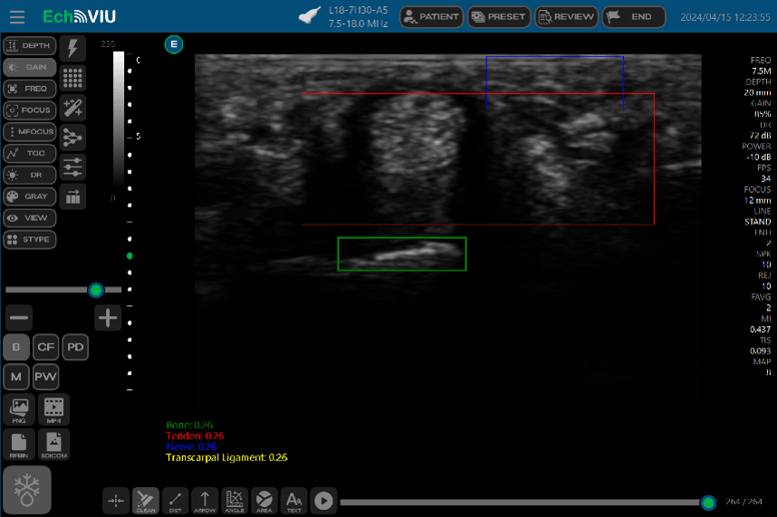
Touchscreen Operation with Integrated Tablet Display
Compatible with Windows tablets, which can be embedded directly into the EchoVIU® unit for intuitive touch-based control.

Smart Applications
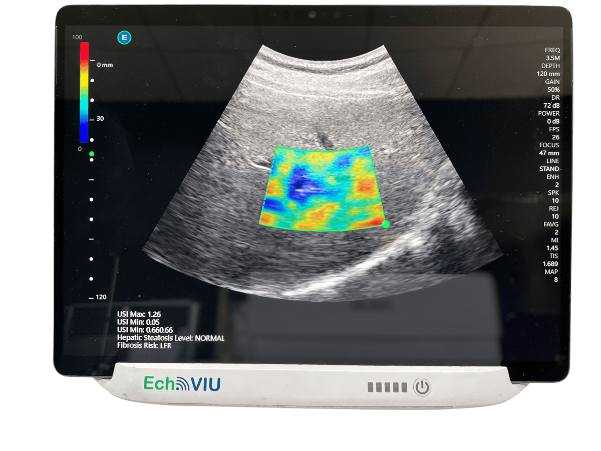
Equipped with AI-enhanced algorithms to effectively solve common clinical challenges faced by physicians.
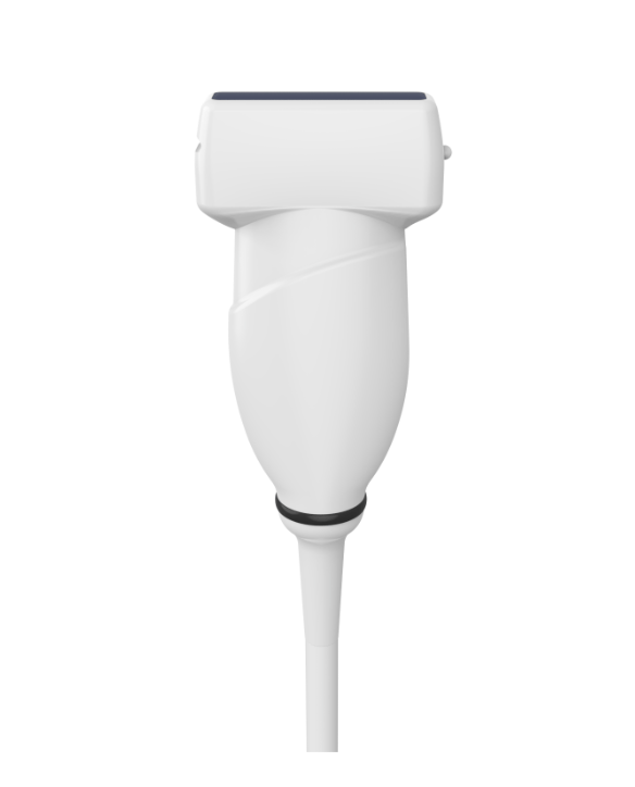
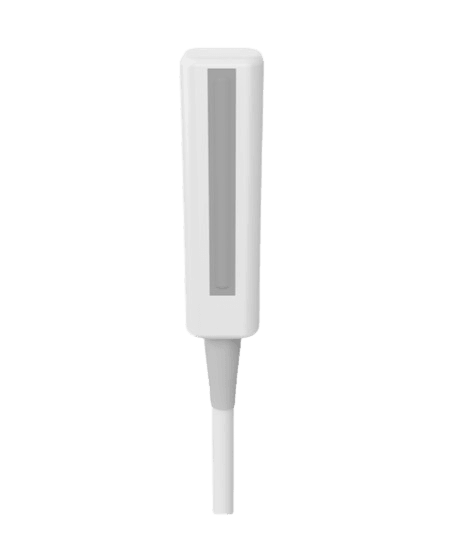
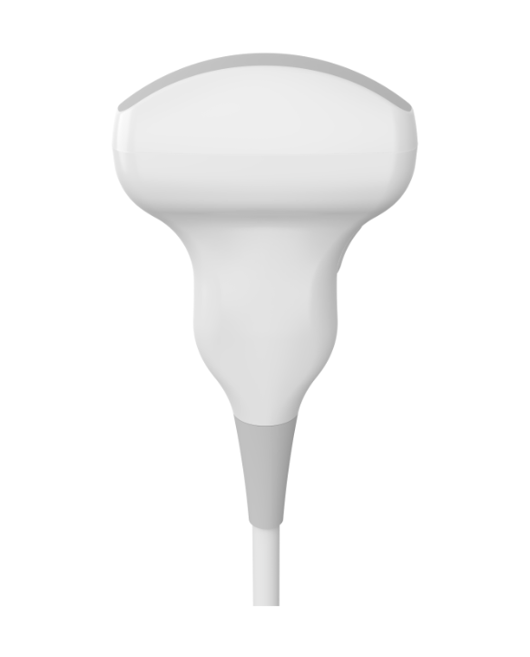
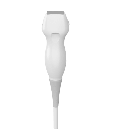
Supported Probes
Linear | Musculoskeletal | Convex | Phased Array

L18-7H40-A5
Type: Linear
Frequency: 7.0 - 18.0 MHz
Field of View:38.4 mm
Applications: Vascular, Small Parts, Musculoskeletal/Nerve
L15-7H40-A5
Type:Linear
Frequency: 7.0 - 15.0 MHz
Field of View:38.4 mm
Element Number: 192
Applications: Vascular, Small Parts, Musculoskeletal/Nerve

L12-5N40-A4
Type: Linear
Frequency: 5.0 - 12.0 MHz
Field of View:38.4 mm
Element Number: 128
Applications: Vascular, Small Parts, Musculoskeletal/Nerve

LF9-5N60-A3
Type: Linear
Frequency: 5.0 - 9.0 MHz
Field of View: 64 mm
Element Number: 128
Application: Musculoskeletal/Nerve

C5-2H60-A5
Type: Convex
Frequency: 2.0 - 5.0 MHz
Field of View: 60.5°
Element Number: 192
Applications: Abdomen, Gynecology, Obstetrics, Pediatrics

P5-1S15-A6
Type: Phased Array
Frequency: 1.0 - 6.0 MHz
Field of View: 90°
Element Number: 64
Applications: Abdomen, Heart
Book a Demo
Please leave your message and we will get back to you as soon as possible!
Contact Person *Contact Email *Contact Phone *Hospital/Clinic/Company NameMessagePlease feel free to leave your messageInterested Product Models *